Aging alters configuration of fast- and slow-twitch fibers associated with muscle fiber denervation.
Title: Slow twitch soleus muscle is not protected from sarcopenia in senescent rats
Journal: Exp Gerontol (2010) 45:662-670
Link: [https://doi.org/10.1016/j.exger.2010.04.001]
Title: Severe atrophy of slow myofibers in aging muscle is concealed by myosin heavy chain co-expression
Journal: Exp Gerontol (2012) 47:913-915
Link: [https://doi.org/10.1016/j.exger.2012.07.013]
Title: Denervation causes fiber atrophy and myosin heavy chain co-expression in senescent skeletal muscle
Journal: PLoS One (2012) 7(1):e29082
Link: https://doi.org/10.1371/journal.pone.0029082
Comments:
Sarcopenia is age-related loss of muscle mass (in skeletal muscle) and motor function and is a slow process caused by multiple factors such as inactivity, anorexia, inflammation, aging, endocrine, and neurodegeneration.
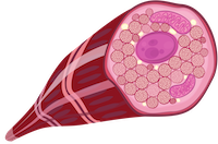
Muscles consist of two types of muscle fibers, slow-twitch and fast-twitch fibers. A current leading hypothesis is that spinal motor neurons innervating fast-twitch fiber are lost, resulting in atrophy (decay of fast-twitch fiber) or transition of fiber type (fast-twitch fiber to slow-twitch fiber) with ages. On the other hand, the slow-twitch fibers have been supposed to be protected from atrophy. Three papers suggest that the slow-twitch fibers are also affected with aging, which contradicts the widely accepted notion.
Current study (Carter et al., 2010 Exp Gerontol) looked into the transition of twitch fibers in soleus which predominantly contains slow-twitch fibers, as well as gastrocnemius, which predominantly contains fast-twitch fibers, by immunohistochemical approach for myosin heavy chain (MHC) isoforms in young adult and senescent (‘aged’) rats. They found a transition from slow-twitch fiber to fast-twitch fiber with age.
Secondary, they addressed how this finding which contradicts the widely accepted notion has been concealed so far (Purves-Smith et al., 2012 Exp Gerontol). By checking co-expression of fast and slow MHC as well as anti-muscle atrophy F-box (biomarker of muscle atrophy) by immunohistochemical approach. They found the increase in co-expression of fast and slow MHC with ages, suggesting that the phenomenon of MHC co-expression has obscured the actual degree of atrophy of MHC slow fibers in the previous studies.
Finally, they addressed the above hypothesis that denervation caused the fiber transition or atrophy (Rowan et al., 2012 PLoS One). Density of motoneurons in lumbar cord decreased with ages. They checked denervated fibers by using Nav1.5 as a marker of muscle fiber denervation. They found that Nav1.5 positive fibers had higher level of ubiquitin ligases (biomarkers of muscle atrophy), suggesting that muscle fiber denervation is associated with the muscle atrophy.
半田 高史
准教授
ドイツ留学から帰国後に広島大学助教として参加し、2025年4月から准教授として活躍。行動中の動物から神経活動を記録する電気生理学を専門とし、その理論的解析にも重点をおいて研究を展開しています。